Home
Kindai-LSU Multiple Sclerosis
Research Group
Myocarditis
Members
Pictures
Publications
Movies
Links
Blog
Lectures
Google
WWW Search Search in this website

Kindai-LSU Multiple Sclerosis Research Team

English 日本語


Kindai-LSU Multiple Sclerosis Research Team Units and Programs
I. What is Multiple Sclerosis?
II. Kindai-LSU Multiple Sclerosis Research Team Overview
III. Neuroimmunology Unit
IV. Vascular Physiology and Microparticle Unit
V. Bioinformatics and Systems Biology Unit
VI. Neurovirology and Neuropathology Unit

I. What is Multiple Sclerosis?

Multiple sclerosis (MS) is an inflammatory demyelinating disease in the central nervous system (CNS= brain and spianal cord). Although the precise cause of MS remains unclear, MS has been proposed to be a disease caused by interactions among autoimmunity (immune responses against our own host tissues), virus infections, and/or genetic factors (Figure 4.1). The autoimmune theory of MS has been supported by clinical findings in which myelin-specific T cells and antibodies have been found in some MS patients. Infiltration of immune cells is observed in the demyelinating lesions. Immunomodulatory drugs, such as glatiramer acetate (copaxone), interferon (IFN)-beta (Avonex, Betaseron, Rebif), or anti-very late antigen (VLA)-4 antibody (Natalizumab, Tysabri), are effective in some patients. Experimentally, sensitization with CNS antigen can induce an autoimmune response, resulting in the inflammatory demyelinating disease, experimental autoimmune (allergic) encephalomyelitis (EAE). Microbial infections, particularly virus infection, have also been associated with MS pathogenesis. Clinically, viruses and antiviral immune responses have been detected in some MS patients. Experimentally, demyelinating diseases can be induced by viruses, such as canine distemper virus and murine hepatitis virus. Among viral models for MS, Theiler’s murine encephalomyelitis virus (TMEV) infection has been widely used since 1975. In the TMEV model, various immune cells play an important role in the pathogenesis of demyelination.

II. Kindai-LSU Multiple Sclerosis Research Team Overview

Multiple sclerosis (MS) is an immune-mediated disease and there are more than 3 million patients in the world, 10,000 patients in Japan, and 4,000 patients with MS in Louisiana. In our Kindai-LSU MS Research Team, several research units have studied pathogenesis of multiple sclerosis. Tsunoda laboratory has studied autoimmune and viral etiologies of MS, while Dr. J Steven Alexander, PhD has investigated roles of microparticle as well as vascular physiology in MS. Drs. Alexander and Tsunoda have collaborated with Dr. Alireza Minagar, MD, Professor, Depatment of Neurology, Louisiana State University Health Sciences Center.
LSU MS Research Team has also contributed to fundrasining and public awareness for MS, joining events such as Walk MS, as LSU Tigers MS Research Team for Walk MS: Shreveport-Bossier City . Walk MS: Shreveport-Bossier City is the first and foremost a fundraising event in North Louisiana. Money raised at this 
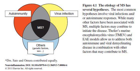
year’s event will support research progress in many areas, moving us closer to our ultimate goal of a world without MS.

Tsunoda laboratory has been supported by Center for Molecular and Tumor Virology, Louisiana State University. The goal of the Center for Molecular and Tumor Virology is to bring together scientists who direct independent but interactive research programs in molecular and tumor virology. Within the CMTV, senior faculty members help to develop the academic careers of the junior level faculty members. The COBRE-supported CMTV includes: The Molecular Analysis Core providing services, reagents, major instrumentation, equipment, and technical support to enhance the research activities of members of the CMTV. 
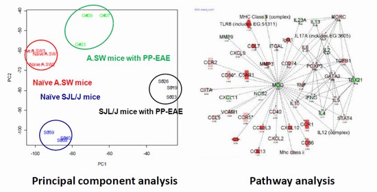
Bioinformatics Core based at the campus of LSU-Shreveport providing a variety of services for data analysis, bioinformatics, and data mining.

III. Neuroimmunology Unit

Tsunoda laboratory has investigated pathogenesis of MS using immunological, virological and pathological methods, while Dr. Matthew Grisham's laboratory has collaborated with Tsunoda laboratory in studying a role of regulatory T (Treg) cells, a new subset of immune cells, in MS (Treg Program). Tsunoda laboratory has also collaborated internationally with Drs. Satoru Takahashi and Keigyo Yo laboratories, University of Tsukuba, Ibaraki, Japan. Using transgenic ("gain of function") method established in Japanese laboratories, Tsunoda laboratory stuied roles of three T helper (Th) cell subsets, Th1, Th2, and Th17 cells in MS model system (Th Transgenic Program).

Dr. Matthew B. Grisham laboratory, Department of Immunology and Molecular Microbiology, Texas Tech University Health Sciences Center

Dr. Grisham's laboratory has collaborated with Tsunoda laboratory in "Treg Program." Matthew B. Grisham laboratory, Department of Immunology and Molecular Microbiology, Texas Tech University Health Sciences Center in Lubbock, Texas. Dr. Grisham established a novel method to expand regulatory T (Treg) cells, which can be used for futture treatment of several immune-mediated diseases, including inflammatory bowel diseases (Ulcerative colitis and Crohn's disease) and multiple sclerosis. Dr. Grisham has collaborated with Tsunoda laboratory. Our team found that Treg can play a different role in the course of our MS model system. Dr. Grisham is currently an Associate Editor of the American Journal of Physiology and Free Radical Biology and Medicine. He has served or is currently serving on the editorial boards of the journals Inflammatory Bowel Disease, the Journal of Immunology and American Journal of Physiology.

Dr. Satoru Takahashi laboratory, Department of Anatomy and Embryology,University of Tsukuba, Japan

Dr. Takahashi's laboratory has collaborated with Tsunoda laboratory in "Th Transgenic Program." An individual's disease sensitivity is determined by T-cell subsets balance. T-cell subsets are divided into T helper (Th)1, Th2, Th17 and regulatory T (Treg) whose transcription factors T-bet, GATA-3, RORgt and FoxP3 are involved in the Th subtype differentiation. Dr. Takahashi's laboratory are interested in analyzing the function of these transcription factors against disease sensitivity determination, mainly T-bet, GATA-3 and RORgt.
Dr. Satoru Takahashi, Professor, University of Tsukuba, hosted a seminar by Ikuo Tsuoda: The 330th Tsukuba Molecular Life Science Seminar. “Resveratrol, a red wine component, exacerbates autoimmune and viral models for multiple sclerosis.” Tsukuba Society for Molecular Medicine/TSMM, University of Tsukuba, Tsukuba, Ibaraki, Japan. September 9, 2011.


IV. Vascular Physiology and Microparticle Unit

Drs. J. Steven Alexander and Alireza Minagar have studied MS as a potential "Vascular disease", investigating vascular physiology as well as microparticle in MS and its model system.

Dr. J. Steven Alexander laboratory, Department of Molecular and Cellular Physiology, LSU Health Sciences Center, Shreverport

Dr. Alexander is an cardiovascular biologist working on how small blood vessels become injured during the process of inflammation. Several diseases including stroke, cancer, Crohn's disease and diabetes are associated with disturbances in how blood vessels restrict exchange of their contents, leading to the development of edema.

Dr. Alireza Minagar laboratory, Department of Neurology, LSU Health Sciences Center, Shreverport

Dr. Alireza Minagar is a board certified neurologist who sub-specializes in clinical neuro-immunology and multiple sclerosis. He sees patients with MS and other inflammatory disorders of the brain and spinal cord at the Neurology Clinic of Louisiana State University Health Sciences Center in Shreveport, Louisiana. Dr. Minagar's research interests involve abnormalities of cerebral endothelial cells in the context of various inflammatory disorders, particularly MS, interactions of cerebral endothelial cells with activated leukocytes, transendothelial migration of leukocytes in MS as well as role of endothelial microparticles in pathogenesis of MS. Dr. Minagar is also involved in a number of clinical research projects related to use of new immuno-modulatory agents for treatment of MS. Dr. Minagar is also interested in the role of various biomarkers in predicting the course of MS and therapeutic response of these patients to immunomodulators.

V. Bioinformatics and Systems Biology Unit

Tsunoda laboratory has collaborated with Drs Marjan Trutshcl and Urska Cvek laboratories, Computer Sciences Department, LSU-Shreveport. Tsunoda laboratory has conducted biological experiments, using microarray, immunological, virological, and pathological methods. The enormous and multiple data obtained in the experiments were processed using multivaliate analyses, for example, principal component analysis and pathway analysis.

Dr. Marjan Trutschl laboratory, Computer Science Department, LSU-Shreverport

Dr. Marjan Trutschl is an Associate Professor of Computer Science, co-Director of the Laboratory for Advanced Biomedical Informatics at LSU Shreveport (LSUS) and Director of the Biomedical Informatics Core at the Center for Molecular and Tumor Virology at the Louisiana State University Health Sciences Center in Shreveport (LSUHSC-S). He received his Sc.D. and M.S. in Computer Science from the University of Massachusetts Lowell (UML).

Dr. Urska Cvek laboratory, Computer Science Department, LSU-Shreverport

Drs. Cvek and Trutschl are collaborating with Tsunoda laboratory to assist with the computational and bioinformatics aspects of the microarray project of MS research models that proposes "1-stage" versus "2-stage" models of Multiple Sclerosis, an immune-mediated disease. This activity is tightly coupled with the field of Systems Biology – information visualization and analysis of large multivariate and high-dimensional data sets, as applied to a broad spectrum of bioinformatics and cheminformatics data sets. Our main role in this collaboration is knowledge discovery from clustering, principal component analysis (PCA), Significance Analysis of Microarrays (SAM), and data exploration to confirm the hypothesis and determine the molecular mechanisms contributing to clinical courses.

Dr. John A. Vanchiere laboraotry, Department of Pediatrics, Louisiana State Univeristy Health Sciences Center


Dr. John A. Vanchiere, MD, PhD, Associate Professor and Chief of Pediatric Infectious Diseases, uses Luminex xMAP technology, which is a particularly powerful platform for simultaneous, “multiplex” detection of proteins in a single biological sample, in contrast to traditional “singleplex” protein detection methods, such as ELISA or Western blotting, Luminex xMAP technology is one of the fastest growing and most respected multiplex technologies and is applied throughout the life sciences, including clinical diagnostics. Collaborating with Dr. Vanchiere's laboratory, we are currently measure 32 cytokines and chemokines from 25-μL samples, usign Cytokine/Chemokine Magnetic Bead Panel (Millipore).

VI. Neurovirology and Neuropathology Unit

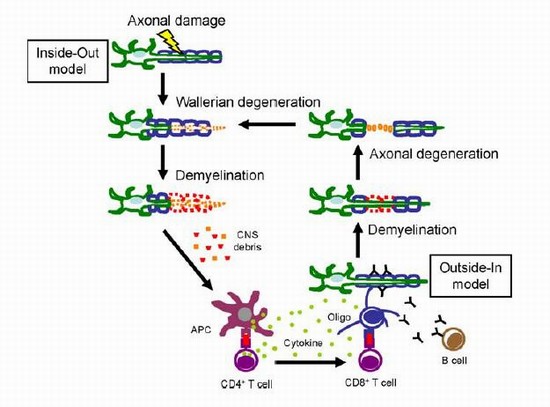
Tsunoda laboratory has also investigated pathogenesis of MS using virological and pathological methods, including laser capture microdissection (LCM). MS is characterized by demyelination and axonal degeneration in the CNS. The primary target in MS has been believed to be either myelin (myelinopathy) itself or oligodendrocytes (oligodendrogliopathy), and axonal degeneration is regarded as a secondary injury to demyelination. In this hypothesis, anti-myelin immune responses or direct virus infections of oligodendrocytes play an effector role in inflammatory demyelination. If inflammation that causes myelin damage is severe, it can secondarily damage axons. Here, the lesion develops from myelin (outside) to axon (inside), hence the name “Outside-In” model. EAE models induced by myelin antigens support the Outside-In model. However, histologic and neuroimaging studies demonstrated that axonal degeneration in the absence of demyelinating lesions occur in some MS patients, which cannot be explained by the classic Outside-In model.

In viral models for MS, including TMEV and canine distemper virus infections, axonal degeneration has been shown to precede demyelination. In addition, EAE has been shown to be induced by axonal antigens, including neurofilament light protein and contactin-2/transiently expressed axonal glycoprotein 1 (TAG-1). Here, the lesion develops from axon (inside) to myelin (outside), “Inside-Out” model. In the Inside-Out model, axonal degeneration can lead to the induction of oligodendrocyte apoptosis, which results in secondary demyelination. Later, degenerated myelin and axonal antigens can be phagocytosed by microglia and macrophages, which can present myelin antigens and induce anti-myelin immune responses. These anti-myelin immune responses, in turn, attack myelin sheaths from the outside. Thus, Outside-In and Inside-Out models can make a vicious cycle, leading to disease progression, unless regulatory mechanisms stop the cascade reaction.

Laser Capture Microdissection (LCM) Core facility, LSU Health Shreveport

Laser capture microdissection (LCM) can be used to separate individual cell types from histology sections. DNA or RNA can then be extracted from these microsections for gene expression analysis. LCM utilizes a low-power infrared laser to melt a special thermoplastic film over the cells or tissues of interest. The PixCell IIe LCM instrument is then used to direct the low-powered infrared laser through the cap to melt the film onto the cells of interest. Dr. Ru Jiang (Department of Microbiology and Immunology, Hutt-Fletcher laboratory, LSU Health Sciences Center, Shreveport) is an expert of LCM and collaborating with our LSU Multiple Sclerosis Research Team to analyze cells from histology sections.


PDF and reprint request Information:

Address: Department of Microbiology
Kindai University Faculty of Medicine
377-2 Ohnohigashi
Osakasayama, Osaka, 589-8511, Japan
Email: itsunoda@hotmail.com or itsunoda@med.kindai.ac.jp
Phone: 81-72-366-0221
Fax: 81-72-366-0206