Myocarditis is an inflammatory disease in the heart and affects about 2 million Americans.
It may occur at any age; infants, immunosuppressed individuals, and pregnant women are particularly vulnerable.
The outcome of patients with myocarditis is poor. As many as 50% of untreated patients die within 2 years of diagnosis.
We do not know what kinds of molecular mechanisms are associated with the pathogenesis of viral myocarditis.
Half of the clinical myocarditis cases in North America are attributable to picornavirus infections.
Myocarditis can be caused by viral infections or immune-mediated
(autoimmune) reactions. Among viruses, picornaviruses are the most common cause of human myocarditis.
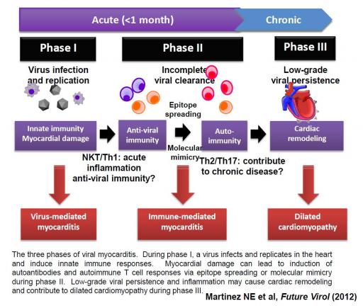
Viral myocarditis has been suggested to develop in three phases. Initial viral replication damages the heart (phase I),
which leads to immune-mediated pathology (phase II), and during the chronic phase (phase III), cardiac remodeling can
lead to dilated cardiomyopathy. However, the precise molecular mechanisms involved in the pathogenesis of viral myocarditis
are unclear. Theiler’s murine encephalomyelitis virus (TMEV) is a member of the genus Cardiovirus, which belongs to the
family Picornaviridae and can cause myocarditis.
Theiler's murine encephalomyelitis virus (TMEV) is a picornavirus and causes inflammatory heart disease,
which is similar to myocarditis found in humans. In our project, we will use the TMEV model and determine temporal
immunological, virological, and pathological responses as well as clinical parameters
(heart specific enzymes and echocardiography).
Among immune cells, we will examine the roles of T helper (Th) cells, regulatory T (Treg) cells and natural killer T (NKT)
cells, which can control microbial infections, including myocarditis. In TMEV infection, Th cells, Treg cells, and NKT cells
have been shown to regulate inflammation in the central nervous system. However, the roles of these immune cells in viral
myocarditis are unknown. We hypothesize that Th cells, Treg cells, and NKT cells play a regulatory role in viral clearance
and disease activities in TMEV-induced myorcarditis. We will determine the roles of these immune cells, adoptive transfer of immune cells, or by depleting these immune cells using antibodies.
We will conduct 1) microarray analyses that will provide comprehensive information of gene expression and 2) a systems
biology approach that can correlate microarray data with immunological (Th, Treg, and NKT cell responses), clinical and
histological findings in the heart in each phase of viral myocarditis. These findings will be associated with microarray gene profiles by
collaborating with computer scientists: Drs. Marjan Trutschl and Urska Cvek, Computer Science Department, LSU-S.
This may lead to the clarification of a molecular mechanism of viral and immune-mediated pathogenesis as well as the
discovery of new biomarkers in myocarditis. Achievement of the aims in this project will allow us to add insight into
possible associations between viral replication, immune responses, disease stages, and cardiac dysfunction and injury to
the current clinical and pathological knowledge of viral myocarditis. In the future, this will drive the development of
personalized interventions of myocarditis, whose treatment can be dependent on the causes (virus and autoimmune), disease
phases, and immunological backgrounds.
Our laboratory studies viral and immune-mediated diseases, including multiple sclerosis and myocarditis.
Myocarditis is inflammatory disease in the heart, whose causes can be viral infection and/or autoimmune responses against heart muscle. We have collaborated an expert in cardiovascular disease research, Dr. J. Steven Alexander, at LSU Health Shreveport. We use cardiomyocyte (heart muscle cell) cell line, HL-1 cells who beat in tissue culture flasks! Using this model system, we are analyzing changes of gene expression patterns in cardiomyocyte, using a microarray analysis.
Our research has been supported by Center for Cardiovascular Diseases and Sciences, Louisiana State University Health Sciences Center. Our Assistant Professors,
Drs. Seiichi Omura and Fumikata Sato have been funded by Malcolm Feist Cardiovascular Research Fellowship grants. The project of
Dr. Omura is "Systems Biology Approach for Molecular Mechanisms in Viral Myocarditis", in which microarray data are analyzed
by bioinformatics approach. The title of Dr. Sato's project is "Regulatory Role of Natural Killer T-cells in Cardiovirus-induced Myocarditis."
In this project, we continue collaboration with Dr. Masaru Taniguchi, Director RIKEN Research Center for Allergy and Immunology, Yokohama, Japan,
studying the immunological role of NKT cells in our myocarditis model.
We analyze cardiac functions of our in vivo model with micro-echocardiography, using the Vevo 770 system in the Institutue for Cardiovascular Disease
and Imaging, Louisiana State University Health Sciencecs Center, Shreveport. Below movies and images were taken, using the Vevo 770 system by Dr. Eiichiro Kawai, MD,
Postdoctoral Fellow, Tsunoda laboratory.
Echocardiography movies
Left: LV-SAX, Short axis view of the left ventricle (LV). There are two papillary muscles seen as two white round structures in the LV cavity.
Right: LV-LAX, Long axis view of the LV. See the apex on the left, and the mitral valve (MV) and the aortic valve on the right.
Pulmonary valve and artery. Pulmonary valve motion.
Other Echocardiography image
Left: LV M-mode. By the M-mode view, We can see the wall movement and measure fraction shortening (FS) and Ejection Fraction (EF). In this case, FS and EF are 0.50 and 0.82 respectively.
Right: Aorta flow. Blood flow of aorta by PW mode.
Left: Aortic arch (AA). Aortic arch and its branch are seen. Pulmonary artery (PA) is seen like as it is surrounded by the Aorta.
Right: Pulmonary artery flow. Pulmonary artery flow by PW mode. Pulmonary regurgitations were detected in some of the cases.
Mitral valve inflow.
MV flow by PW mode. There are two peaks of the flow (E wave and A wave). These peaks are, however, fused and unclear when the heart rate was high.
Contact Information:
Address: Department of Microbiology
Kindai University Faculty of Medicine
1-14-1 Miharadai, Minami-ku
Sakai, Osaka 590-0197 Japan
Email: itsunoda@hotmail.com or itsunoda@med.kindai.ac.jp
Phone: +81-72-288-7222 (ext. 5040)
Fax: +81-72-298-1500